Javascript is not enabled.

Javascript must be enabled to use this site. Please enable Javascript in your browser and try again.

Skip to content
Content starts here
CLOSE ×
Search
CLOSE ×
Search
Leaving AARP.org Website

You are now leaving AARP.org and going to a website that is not operated by AARP. A different privacy policy and terms of service will apply.

What is Diabetic Retinopathy: Symptoms, Stages and Treatment

What to know about diabetes’ association with vision loss


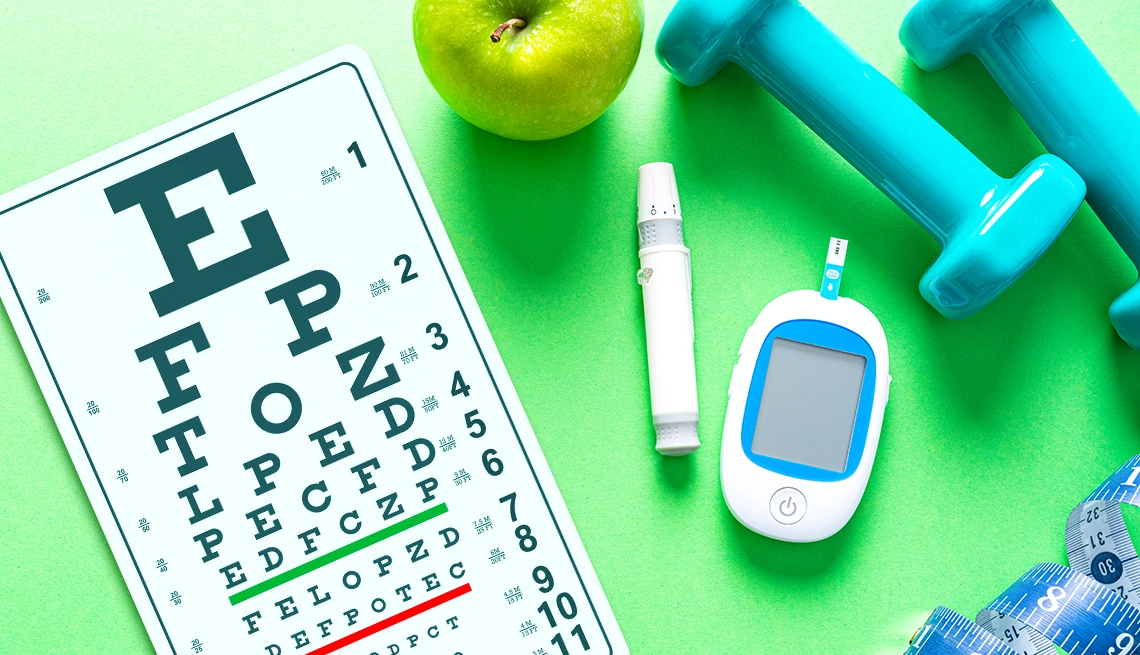
an eye test, a diabetes test, an apple with blue weights
Photo Collage: AARP; (Source: Getty Images (2))

Not paying attention to your blood sugar can lead to a lot more than some high digits on your bathroom scale. There’s a laundry list of health complications that come from lofty glucose levels — among them, nerve damage in your hands and feet, kidney damage, heart disease and stroke. And then there are your eyes.

What is diabetic retinopathy?

People who have diabetes — Type 1 or Type 2 — are at risk for diabetic retinopathy, a condition where consistently high blood-sugar levels damage the blood vessels in the retina, the thin layer of tissue at the back of the eye that’s essential for maintaining vision.

In fact, diabetic retinopathy is the leading common cause of vision loss among people with diabetes and the most frequent cause of new cases of blindness among adults aged 20 to 74, a 2023 study found.

What causes diabetic retinopathy?

The onset of diabetic retinopathy largely depends on the type of diabetes a person has and how long they’ve had it.

For example, people with type 1 diabetes are often diagnosed with diabetes at a younger age because their symptoms tend to be more severe. While they’re typically diagnosed early, it can still take years for diabetic retinopathy to develop.

In contrast, type 2 diabetes — more common among older adults — can go undetected for years. Many people may not realize they have diabetes, especially if they haven’t seen a doctor in a while and feel fine. By the time they’re diagnosed, diabetic retinopathy may have already progressed to a more advanced stage.

“If you get a new diagnosis of diabetes, especially type 2, it’s important to go to your eye doctor and get it checked,” says Lisa Olmos de Koo, professor of ophthalmology at the University of Washington School of Medicine.

Stages and symptoms of diabetic retinopathy

In the early stages of diabetic retinopathy, you might not even know you have it. But as it worsens, your vision takes a hit. It may fluctuate between clear and blurry. You may get floaters (spots or dark strings in your vision), poor night vision, dark or empty areas in your vision, or colors that appear faded. Left unchecked, it can lead to permanent vision loss.

The disease is categorized in two phases:

Nonproliferative diabetic retinopathy  (NPDR)

NPDR is the earliest stage and most common form of diabetic retinopathy. In this stage, tiny blood vessels in the retina are damaged, leading to a variety of issues. Blood spots, known as dot blot hemorrhages, and dilated blood vessels, called microaneurysms, are common signs. Additionally, cotton wool spots — areas with reduced blood flow—can develop, along with fatty deposits known as exudates.

These changes can blur your vision, and sometimes lead to swelling of the retina (macular edema), where fluid accumulates in the center of the retina, worsening vision in a way that cannot be corrected with glasses.

If untreated, chronic macular edema can result in irreversible damage to your sight.

Proliferative diabetic retinopathy (PDR)

PDR is the most severe stage of diabetic retinopathy. Here, the damaged blood vessels in the retina have closed off, cutting off oxygen supply to the retina. In response, the body grows new blood vessels along the surface of the retina to try to restore nourishment. However, these new vessels are fragile and can easily burst with minor activities. When they bleed, they may fill the eye with blood, creating further complications and increasing the risk of retinal detachment as they pull on the retina. What’s more, scar tissue may form around these new blood vessels, which can contract and pull on the retina, potentially causing retinal detachment.

“Sometimes you can have proliferative diabetic retinopathy and not even know it, says Olmos de Koo. “The central vision might be good, but you might have new blood vessels growing in the periphery or on the edges of the retina that haven't yet bled or tugged.”

What does diabetic retinopathy look like in the eye?

If you notice new eye floaters — tiny black specks or strands in your vision — it’s important to schedule a dilated eye exam, even if you don’t have diabetes. Only in very rare and severe cases can signs of diabetic retinopathy be visible by looking in the mirror.

A dilated eye exam allows the doctor to thoroughly inspect the retina — the light-sensitive tissue lining the inner eyewall, which contains delicate blood vessels. During the exam, your eye doctor will check for signs of retinopathy, including blood spots, fatty deposits, swelling, or the growth of new, fragile blood vessels.

Diabetic retinopathy maintenance and prevention

Even if you don’t have diabetes, adults age 50 and older should check in with their primary care provider annually to rule out a diabetes diagnosis.

“Sometimes, diabetes can creep up on you. You may feel fine and not be aware, causing damage in your eyes,” says Olmos de Koo.

For those with diabetes, prevention starts with managing blood sugar levels. It's essential to work with your primary care provider or endocrinologist to discuss medications, lifestyle changes, and dietary adjustments that can help control blood sugar.

“Even with the best ophthalmology treatments that we can provide, if the blood sugar remains high, a lot of times we’re not going to make a lot of leeway,” says Olmos de Koo.

If you have mild or moderate NPDR, immediate treatment may not be necessary, but your ophthalmologist will monitor your eyes closely. Keeping your blood sugar in check can usually halt the progression and curb vision loss, and sometimes can even restore some of your vision.

An ongoing study since 1984 has shown that maintaining lower A1c levels reduces the likelihood of developing vision-threatening retinopathy.

It’s important that patients with Type 1 diabetes have annual screenings five years after the onset of their disease. Patients with Type 2 should have a prompt examination at the time of diagnosis, and at least a yearly examination, says Rahul Khurana, M.D., an associate clinical professor in ophthalmology at the University of California San Francisco Medical Center and President of the Northern California Retina Vitreous Associates.

“Nearly 40 percent of Americans who have diabetes are not getting a dilated eye examination, and that’s a big problem,” Khurana adds. “We know that 95 percent of vision loss from diabetes can be prevented, but that can only happen if we examine patients early.”

 Monitoring blood pressure and cholesterol levels is crucial for protecting your vision. High blood pressure can exacerbate damage to the fragile blood vessels in the retina, while elevated cholesterol can lead to fatty deposits that further harm these delicate vessels. Together, these factors can accelerate the progression of diabetic retinopathy and worsen vision loss.

“Imagine a garden hose that is weak and leaking,” explains Omesh P. Gupta, M.D., an ophthalmologist and retina specialist at Wills Eye Hospital in Philadelphia. “When your blood pressure is high, it’s like turning up the faucet, so the hose starts leaking even more severely.”

Treatment options for diabetic retinopathy

To halt or even reverse diabetic retinopathy, treatment options depend on the symptoms and underlying cause of vision loss.

For diabetic macular edema (swelling of the retina that blurs vision in ways that glasses can’t correct), treatments typically involve eye injections using a small needle. There are two primary types of medications:

  • Anti-vascular endothelial growth factor (VEGF): This treatment makes blood vessels less leaky and can shrink new, abnormal blood vessels.
  • Steroids: These help reduce swelling in the retina.

Not only can we prevent patients from losing their vision, some patients can actually improve their vision,” says Khurana. Anti- VEGF injections are also used to prevent NPDR from progressing to more advanced stages.

For new blood vessel growth, laser treatments called pan-retinal photocoagulation (PRP) are available. This involves making thousands of tiny laser burns to shrink the abnormal blood vessels and stabilize the retina, reducing the risk of vision loss. Typically, this treatment is done in one session at an ophthalmologist’s office or eye clinic, although some patients may require additional sessions. Blurry vision is expected for about a day after the procedure.

In severe cases, such as retinal detachment or significant bleeding, retinal surgery (vitrectomy) may be necessary. This procedure clears out the blood. If there is a detachment, a bubble of silicone oil or gas can be used to hold the retina flat. Laser can be applied to any retinal tears and to perform PRP to shrink and stabilize new blood vessels.

At the University of Washington, Olmos de Koo is collaborating with primary investigator Dr. Kasra Rezaei on a study investigating oral medication as a treatment option for diabetic retinopathy. The study focuses on fenofibrate, a drug traditionally used to lower fat in the bloodstream. Another imaging study at UW is exploring non-invasive methods to analyze blood flow in the eye without the need for injected dye — an important step since diabetic retinopathy is largely caused by inadequate blood flow to the retina.

generic-video-poster

Unlock Access to AARP Members Edition

Join AARP to Continue

Already a Member?

Eye Health: Causes and Symptoms

Knowing early warning signs may prevent permanent damage